New functionalities in MRI scanners
Modern magnetic resonance imaging (MRI) scanners have a static magnetic field 60,000 times stronger than that of the Earth.
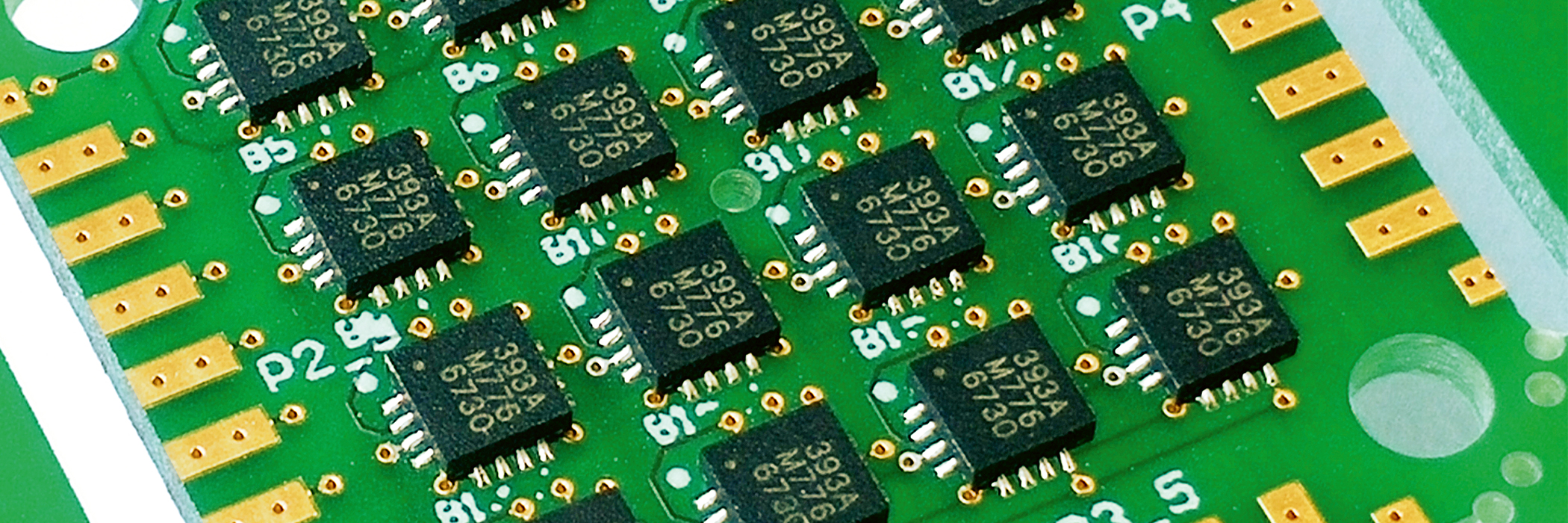
The imaging process is based on small deviations in this field. These ‘magnetic field gradients’ are used to locate and measure resonance signals, making it possible to determine the composition of structures in the human body. Researchers at the Institute for Medical Engineering and Medical Informatics have developed a method using nine sensors to measure these magnetic field gradients extremely precisely. The result is a real time 3D map showing the strength and orientation of the gradients inside the MRI tube at each individual measured point. This new procedure greatly expands the range of MRI applications. The magnetic field sensors are only a few micrometres in size so they can be mounted on ECG electrodes or in surgical tools to track their position in the MRI tube in real time. The concept can also characterise gradient quality: the system is fast enough to detect fields lasting milliseconds, which can help quality control and the characterisation of new magnet designs.
