3D-CT scans
X-ray CT Scanning
By Niko Plath
3-dimensional (3D) computed tomography (CT) is an imaging X-ray technique that generates a 3D radiographic image of an object. For the measurement, the object is placed on a rotating table between an X-ray source and a detector. During the measurement, the object is rotated step by step through 360° and more than 1600 individual X-ray images are recorded. These are processed creating a volume data set as the basis for the examination of the object. This data set can be used to answer various questions about the object.
The method is particularly suitable for the examination of musical instruments. With this nondestructive technique the important, but mostly inaccessible inner volumes of the instruments can be made visible and explored. In the 3D digital representation, measurements can be made with high accuracy and in all possible directions at any desired location. The orthogonal views allow insights into the wood structure, show otherwise invisible repairs, cracks, damages or tool marks. Last but not least, the data can be used to produce exact 3D copies, e.g. by additive processes such as 3D printing.
The investigations of the fagottini were carried out at the Fraunhofer Development Center for X-ray Technology (EZRT) in Fürth/Bavaria. By utilizing a high-resolution CT setup, which is mainly used in industrial material testing, a spatial resolution of 96 μm has been achieved. This means that measurements in a volume data set can be performed or reproductions can be made from a surface data set with a precision of 0.1 mm.
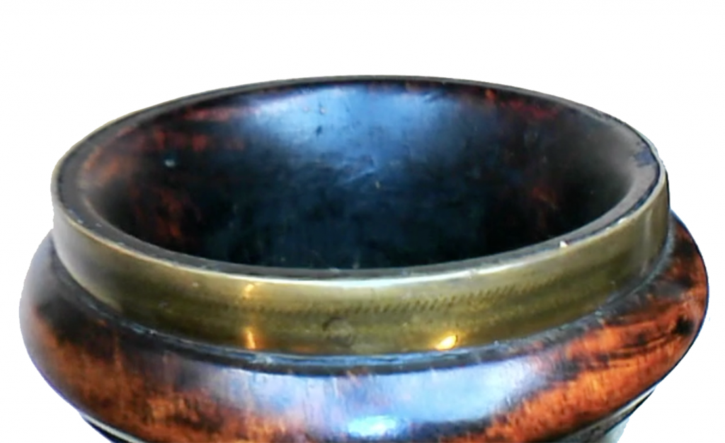
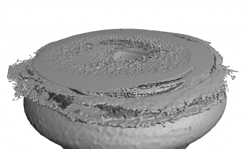
One of the challenges of the method is that image errors (so-called artefacts) can occur when objects are examined with components of different densities, such as wood and metal. The formation of these artefacts is physically caused by the poly-chromatic spectrum and the deflection of the photons by the dense materials (so-called beam hardening effect) and can unfortunately not be avoided. In the case of an old musical instrument, it is not always possible to remove all metal parts before measurement for conservation reasons. Therefore, the resulting image errors must be processed and removed in a complex work process after the measurement.
Post-Processing
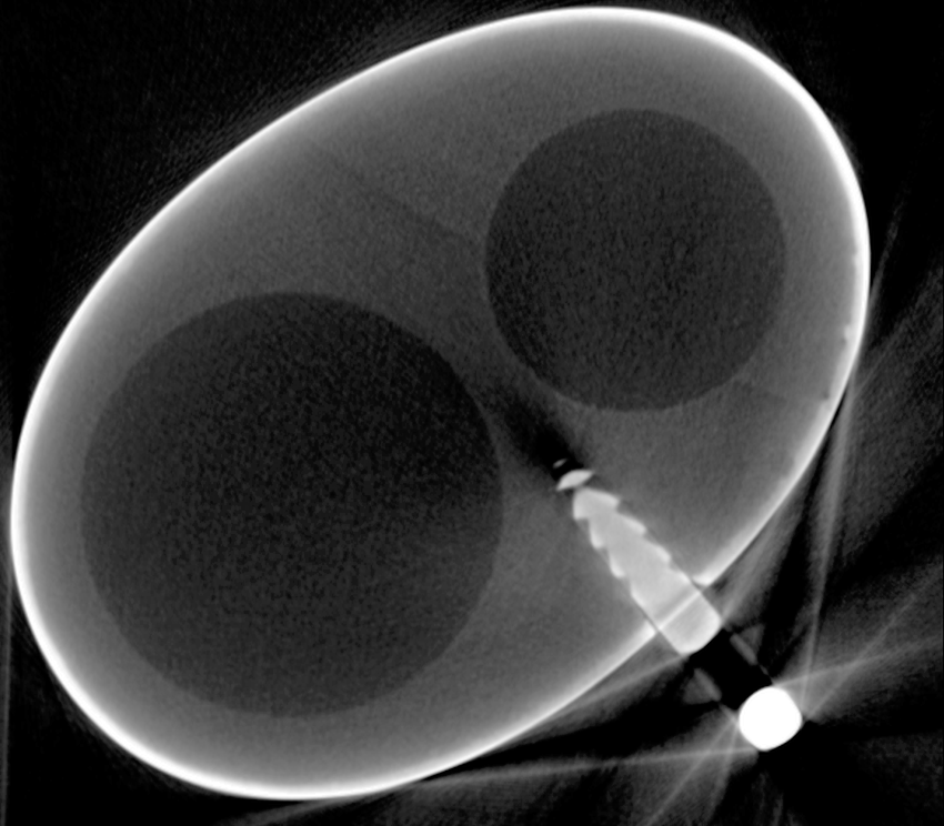
An X-ray CT measurement results in a discrete 3D volume with densities represented by gray values mostly stored in the medical file format DICOM. Figure 1 shows an exemplary cross section. By classification and segmentation, 3D image representations of the included objects can be derived from the CT measurement volume. Segmentation allows to distinguish and extract sub-structures with homogeneous properties from a heterogeneous object. It is widely used in medical applications to discriminate tissues or to generate content-related regions in machine vision. Each voxel (a pixel in 3D space) is allocated to exactly one of the defined sub-volumes, therefore, different sub-volumes cannot overlap, which is crucial for subsequent applications like physical modeling or rapid prototyping. Since sub-volumes are generated according to their homogeneous properties, the respective surface data adequately describes a body. This leads to a massive reduction of data. Handling, therefore, is simplified (3D meshes of instrument parts can easily be sent per email) and individual sections can be stored in databases for subsequent research tasks. For the exact determination of the surface, a scan with as less noise and other image errors (e.g. metal artefacts) as possible is necessary. Figures 2a/b depict the result of the so-called beam-hardening effect on the resulting images. However, even for an excellent scan the exported surface meshes may have diverse defects, e.g. self-intersecting faces, non-manifold edges or unreferenced vertices. A reliable way to obtain a clean and watertight mesh is a Poisson Surface Reconstruction but note that since this is actually a re-meshing of the polygon surface, individual sub-volumes could overlap afterwards.
Printing
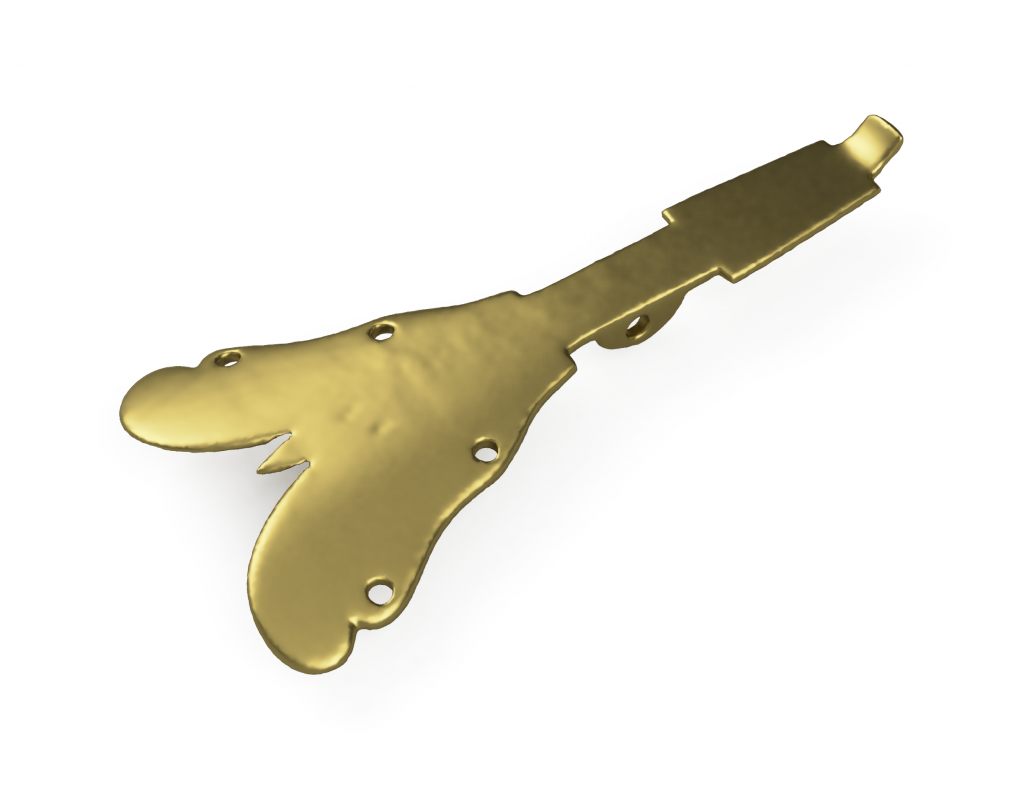
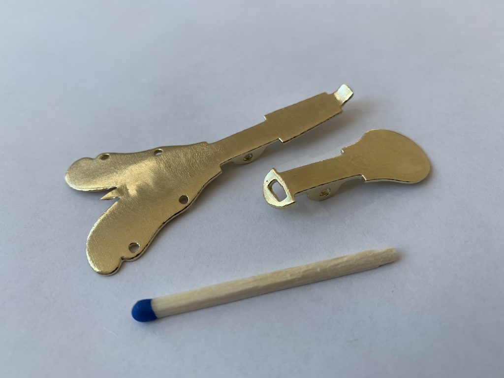
Since historical instruments are in many cases not in playable condition, exact replications are often manufactured and subsequently modified to playable condition. Using extracted surface data of CT scans, whole instruments can be reproduced with high geometric precision as well as missing parts for restoration purposes. Manufacture may be obtained by additive (3D printing) or subtractive methods (CNC milling). Several consumer 3D printers based on fused deposit modeling (FDM) can achieve a vertical spatial resolution of 100 μm, printers based on stereolithography (SLA) realize comparable resolutions in all axes. The obtained 3D models can also be utilized for metal casting. A negative mold is produced based on the surface mesh of the object, which can then be filled with various metals in liquid form. See Figures 3a/b for a comparison between a rendered model and cast brass part.