With the newly acquired Horiba XploRA™ PLUS Raman microspectroscope, plastics can be examined with high precision in three dimensions.

Raman spectroscopy is a molecular spectroscopic technique that uses the interaction of light with matter to gain insight into the properties of a material. The information provided by Raman spectroscopy results from a light scattering process, while in comparison, infrared spectroscopy is based on the absorption of light. Raman spectroscopy can contribute to the understanding of a reaction based on additional information about intra- and intermolecular vibrations. Raman spectroscopy, which is equivalent to infrared spectroscopy, works with spectra that are characteristic of the specific vibrations of a molecule and are valuable for identifying a substance ("chemical fingerprint"). However, Raman spectroscopy can provide additional information about low-frequency modes and vibrations that give insight into the crystal lattice and molecular backbone structure.
The new Horiba XploRA™ PLUS is equipped with three laser types: 532 nm, 638 nm, and 785 nm. The spatial resolution in the x- and y-direction is < 500 nm, and in the z-direction < 2 μm, showing clear advantages over infrared spectroscopy. Standard objectives with magnification 5x, 10x, and 100x as well as a 60x objective for water immersion are available. The Raman microspectroscope allows the user to change samples very easily, as the mode of operation is comparable to a confocal microscope. The samples do not require any specific preparation and can be used immediately. It has no influence on whether a ground sample or fragments of a component are used. Another advantage is the coupling of the evaluation software with existing material libraries, which can be used to characterise a sample within a short time using reference spectra. In the following, we will present two practical applications of how the Raman microspectroscope is already being used in our institute.
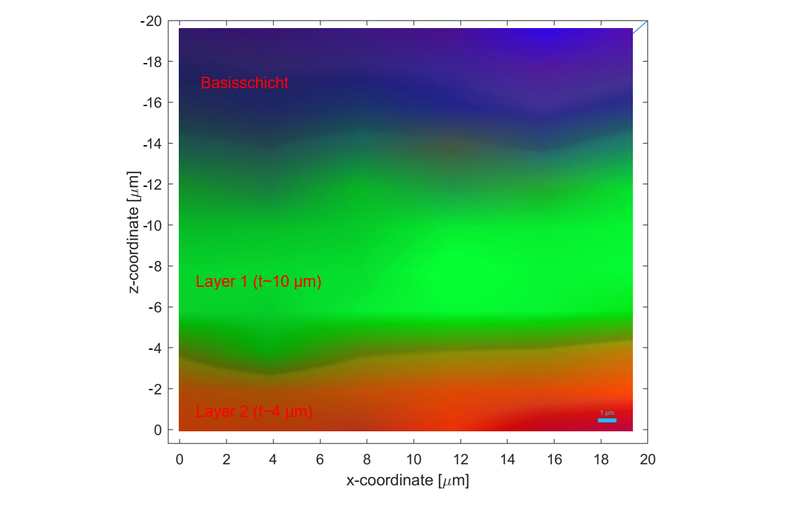
Characterisation of multilayer systems
Neben der normalen Charakterisierung eines Polymers gibt es auch komplexe Methoden, welche genutzt werden können, um beispielsweise die Dicken einer mehrschichtigen Folie zu bestimmen. Hier ist anzumerken, dass es mit dem Raman Mikrospektroskop möglich ist, in Dickenrichtung Messungen durchzuführen, ohne dafür die Probe zu zerstören beziehungsweise aufzutrennen. Dies ist stark abhängig von der Transparenz, der Dicke sowie der molekularen Struktur des Materials. Nachfolgend ein Beispiel einer Messung in Dickenrichtung, wobei mittels mehrerer Spektren in x- sowie z-Richtung ein farbkodiertes Bild generiert wurde, welches verdeutlicht, an welchem Punkt die neue Schicht beginnt.
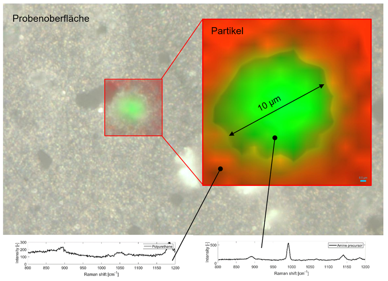
Investigation of filler content in polymers
Another application is the investigation of filler fractions and particles in a polymer matrix. There are two different approaches:
- On the one hand, it is possible to manually select an area which is then measured step by step by the Raman microspectroscope, and based on the change in the spectra, a particle can in turn be colour-coded (see image below). This provides spatially resolved information about the composition, size, and orientation of the particle.
- On the other hand, there is the possibility of using existing software, the "ParticleFinder" from Horiba, which automatically evaluates a selected area based on an algorithm. In this way, a statistical distribution of the particles present in terms of composition, size, etc. can be determined.