Füllung von Knochendefekten mit offenporösen Gitterstrukturen
Die Behandlung grosser Knochendefekte stellt nach wie vor eine grosse Herausforderung in der orthopädischen und kranio-maxillofazialen Chirurgie dar. Eine mögliche Lösung ist die Entwicklung personalisierter poröser Implantate auf Titanbasis, die alle mechanischen Anforderungen mit einem Minimum an Titan und einem Maximum an osteopromotiven Eigenschaften erfüllen.
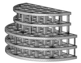
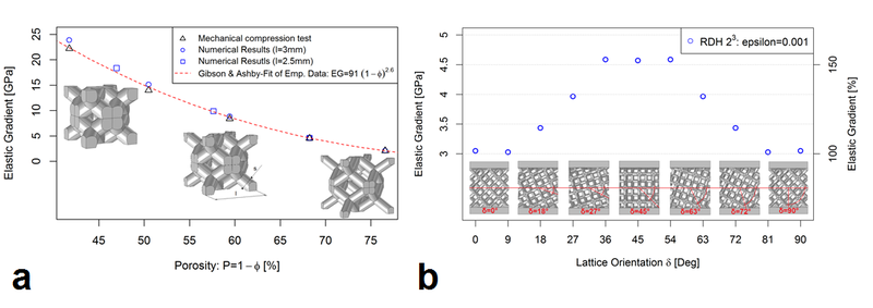
In diesem Projekt haben wir spezifische Designs von Einheitszellen entwickelt, um Knochendefekte mit einer offenporösen Gitterstruktur zu füllen. Die mechanische Reaktion des Gerüsts hängt von der gewählten Gitterarchitektur ab. Dies ist von grosser Bedeutung für die Nachahmung menschlicher Knochen durch Titan-Gerüste, um die Stressabschirmung zu reduzieren.
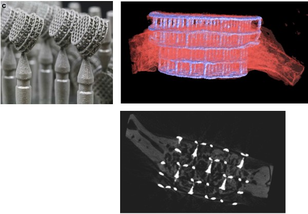
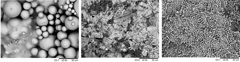
Wir druckten die entworfenen Titan-Gerüste mittels selektivem Laserschmelzen in 3D und implantierten sie anschliessend in Schädeldefekte von Kaninchen, um die Knochenbildung und Osseointegration zu untersuchen. Wir stellten signifikante Unterschiede zwischen den mit Implantaten gefüllten Defekten und den unbehandelten Defekten fest.
Die Studien zielten ferner darauf ab, SLM anzuwenden, das ein hohes Mass an mikroarchitektonischer Freiheit bei der Erzeugung von Gitterstrukturen ermöglicht, und den optimalen Abstand zwischen den Stäben sowie den optimalen Durchmesser der Stäbe für die Osteokonduktion (Einwachsen von Knochen in Gerüste) und die Knochenregeneration zu bestimmen. Für die biologische Auswertung setzten wir verschiedene SLM-gefertigte Titanimplantate in die Schädeldecke von Kaninchen ein und bestimmten nach vier Wochen Heilungszeit die neue Knochenbildung und die Defektüberbrückung. Um die 3D-Gerüstarchitektur mit den biologischen Messwerten zu verknüpfen, bestimmten wir das Einwachsen von Knochen, den Kontakt zwischen Knochen und Implantat sowie die Defektüberbrückung von nicht kritischen Defekten im Schädelknochen von Kaninchen. Darüber hinaus ermittelten wir die optimale Mikroarchitektur für die Osteokonduktion und bestimmten die Druckfestigkeit und den Elastizitätsmodul der ausgewählten Architekturen.
Media
Publikationen
- Osteoconductive Lattice Microarchitecture for Optimized Bone Regeneration
- Mechanical anisotropy of titanium scaffolds
- Stiffness-anisotropy of porous implant geometries
- Influence of Microarchitecture on Osteoconduction and Mechanics of Porous Titanium Scaffolds Generated by Selective Laser Melting
- Lattice Microarchitecture for Bone Tissue Engineering from Calcium Phosphate Compared to Titanium
Projektdetails
- Typ
- Forschungsprojekt
- Forschungsfeld
- Funktionale Materialien und Oberflächen
- Hochschule/Institut
- Hochschule für Life Sciences FHNW / Institut für Medizintechnik und Medizininformatik
- Partner
- Universitätsspital Zürich
- Förderung
- AOCMF, SNSF
Kontakt
Prof. Dr. Michael de Wild
- Telefon
- +41 61 228 56 49
- michael.dewild@fhnw.ch